


Service hotline
+86 18518316054
 Current location : Home page > Resources > Papers > Facile synthesis of visible light α-Fe2O3/BiOBr composite with highly photocatalytic performance
Xiaoju Wena , Chang Zhanga , Chenggang Niua *, Lei Zhanga , Dawei Huangb *, Xiaoyu Wang a, Xuegang Zhang a, Guangming Zenga a College of Environmental Science Engineering, Key Laboratory of Environmental Biology Pollution Control, Ministry of Education, 6 Hunan University, Changsha 410082, China b South China Institute of Environmental Sciences, Ministry of 8 Environmental Protection of PRC, Guangzhou 510655, China
Abstract
Novel α-Fe2O3/BiOBr composites were synthesized by a simple in hydrolysis method for the first time, and were fully characterized by X-ray diffraction patterns (XRD), transmission electron microscopy (TEM), high-resolution transmission electron microscopy (HRTEM) and UV–vis diffuse reflectance spectra (DRS). The α-Fe2O3/BiOBr composite showed much higher visible-light-driven (VLD) photocatalytic activity than pure α-Fe2O3 and BiOBr for rhodamine B (RhB) degradation. Specifically, the 10Fe/100Bi composite showed the highest photocatalytic activity for the degradation of dyes under the visible light irradiation. The stability of the photocatalytic is found to be satisfying, which makes it potential in practical application. In addition, the active species trapping experiments showed that •O2− were the dominant reactive species. Subsequently, a possible degradation mechanism is proposed. The high photocatalytic activity could be attributed to the enhanced light absorption and the improved separation of photogenerated charge carriers, due to form a p-n heterojunction between α-Fe2O3 and BiOBr.
1 Introduction
Semiconductor photocatalysis offers an effective strategy for organic 33 pollutant remediation and water splitting.1-4TiO2, the key photocatalyst for convenient wastewater treatment and other essential cleaning processes, Nevertheless, suffers from problems such as ineffective sunlight capture and conversion due to its wide band gap(about 3.2eV).5-7 Thus, it is urgent and necessary to develop highly efficient visible-light driven photocatalysts for pollutants degradation.
α-Fe2O3, a n-type semiconductor with a narrow band gap of 2.2eV, has been intensely used as a material for electrochemical electrode, photocatalyst, and gas sensors due to its sufficient stability and absorption capability in the visible light region. Its abundant raw materials, environmentally friendliness, excellent conductivity (high discharge and charge current rates) and stability are preferable features for large-scale production and applications. Because of the distinct properties of α-Fe2O3, it not only improved the separation and transport of photo carriers, but also might cause a higher conduction band position indicating a stronger reductive power.8, Recently, the hierarchically structured α-Fe2O3/Bi2WO6 was reported and exhibited much enhanced photocatalytic activity in degradation of acid red G dye and RhB dye under visible-light irradiation; a Fe2O3/BiOCl p-n heterojunctions with high photocatalytic activity for the degradation of mixture dyes under visible light was synthesized. Therefore, the combining of α-Fe2O3 and another semiconductor with a suitable valence band and conduction band positions is a promising strategy.
Up to now, Bismuth-based photocatalytic materials such as Bi2O3, BiOBr, BiVO4, BiOCl, BiOI etc.19-22 have aroused great interest in the scientific community due to their intriguing electronic structures. Among them, BiOBr has stimulated researcher intensive interest for its visible light response and higher stability.23, 24 Although pure BiOBr samples was synthesized by different methods with various architectures,25, 26 it is still imperative to further improve its photocatalytic activity for practical applications. Fabrication of a heterojunctions composite containing BiOBr has been turned out an attractive strategy for enhancing the photocatalytic activity of BiOBr. Recently, BiOBr–ZnFe2O4 heterojunctions were reported and exhibited excellent ability for the degradation of organic dyes under visible light irradiation.27 what’s more, enhanced photocatalytic activities have also been studied over BiOBr– AgBr and BiOBr–Bismuth oxyhydrate composite materials.28, 29These results motivated us to design α-Fe2O3/BiOBr heterojunctions that are expected to be ideal for improving photocatalytic activity.
Herein, a series of α-Fe2O3/BiOBr composites were prepared by a simple in hydrolysis method. We have investigated the activities of the catalysts at different molar ratios of Fe2O3 to BiOBr. Furthermore, the mechanism of the enhanced photocatalytic efficiency of the composite was discussed.
2 Experimental
2.1 Fe(OH)3 precursor preparation
All the chemicals are of analytical reagent grade and were used without further purification. Distilled water was used in all our experiments. The α-Fe2O3/BiOBr composite was synthesized via a hydrothermal process, using Fe(OH)3 as precursor. Briefly, Fe(OH)3 precursor was firstly obtained by mixing Fe(NO3)3·5H2O and 0.1M NaOH aqueous solutions. After being rinsed with deionized water, the precipitation was sonicated for 30 min to disperse in deionized water completely.
2.2 α-Fe2O3/BiOBr composite sample preparation
The α-Fe2O3/BiOBr composite was synthesized via a hydrothermal 88 process, using Fe(OH)3 as precursor. In a typical procedure, 2mmol Bi(NO3)3·5H2O and 2mmol KBr were dissolved in 30 mL and 10mL deionized water, respectively. After that, these two solutions were mixed together and stirred for 30 min until a white suspension was formed. Then different volume (5.0, 10.0, 15.0 and 20.0 mL) of the Fe(OH)3 suspension was added dropwise to the mixture in order to obtain the dark red suspension with a Fe/Bi molar ratio of 0.05, 0.10, 0.15 and 0.20, respectively. After stirring for 30 min, the reaction mixture was transferred into a 50 mL Teflon-lined stainless steel autoclave, kept at 160℃ for 10 h, followed by natural cooling to room temperature. The product was collected by centrifugation, washed thoroughly with deionized water and alcohol several times, and then dried at 60 ℃ overnight. According to Fe/Bi molar ratio, the as-prepared composite photocatalysts were marked as 5Fe/100Bi, 10Fe/100Bi, 15Fe/100Bi, and 20Fe/100Bi, respectively.
For comparison, the pure BiOBr sample and α-Fe2O3 was also prepared via the same hydrothermal process as described above.
2.3 Characterization
The phase structures of the products were evaluated by X-ray diffraction (XRD) analysis at room temperature on a Bruker D8-advance X-ray diffractometer with Cu Kα radiation (λ = 0.15406 nm). The morphologies and microstructures of the products were obtained by a FESEM-4800 field emission scanning electron microscope (SEM, Hitachi) with 5.0 kV scanning voltages. Transmission electron microscopy (TEM) and high-resolution transmission electron microscopy (HRTEM) images were aimed at further analyzing the morphology and crystallinity of the products on a transmission electron microscope (TEM, FEI Tecnai G20) with an accelerating voltage of 200 kV. Ultraviolet visible (UV-vis) diffuse reflection spectra were examined on a UV-vis spectrophotometer (Hitachi U-4100) in the range of 200 to 800 nm with BaSO4 as a reference.
2.4 photodegradation experiment
The photocatalytic activities of the samples were evaluated by the degradation of RhB aqueous solution under visible light irradiation. A 300 W Xe lamp (CEL-HXF300, Beijing) lamp with a 420 nm cutoff filter was used as the light source. The experiments were performed
 at room temperature as follows: 0.10 g of the as prepared catalyst was added into 100 mL RhB aqueous solutions (20 mg/L). Prior to illumination, thesuspensions were stirred for 30 min in the dark to reach an adsorption– desorption equilibrium of dye molecules on the surface of the photocatalyst. Afterward the suspension was exposed to visible light irradiation while the temperature of the solution was kept unchanged by cooling water. At certain time intervals, 4 mL suspensions were sampled and centrifuged by TGL-16G centrifuge (Shanghai Anting Scientific Instrument Factory, China) at 8000 rpm for 5 min to remove the remaining particles. The upper clear liquid was analyzed by recording the maximum absorption band (553 nm for RhB) and UV–vis spectra of dyes using an UV–vis spectrophotometer (Shimadzu 2550, Japan).
The stability of the α-Fe2O3/BiOBr composite catalyst was evaluated by reusing the catalyst for four runs for the decomposition of RhB under the same conditions. After each run, the catalyst was separated by a simple precipitation procedure and was reused.
3. Result and discussion
3.1. Characterization of samples
3.1.1. XRD analysis
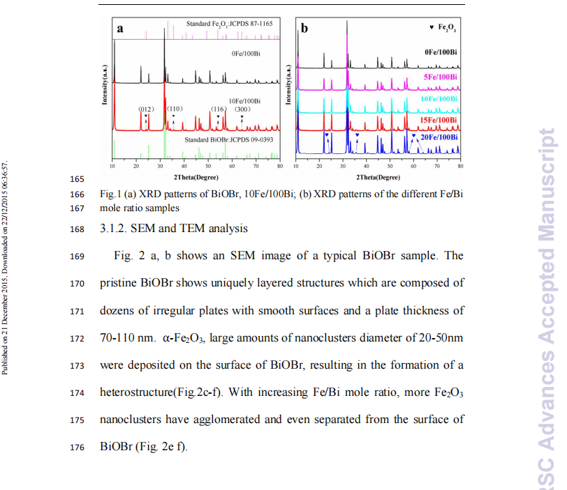
The crystal structure of the products was investigated by the XRD method, as shown in Fig. 1a. As can be seen, all the characteristic peaks in Fig. 1 could be indexed to the tetragonal phase of BiOBr (JCPDS card No. 09-0393) Compared with the pure BiOBr, some diffraction peaks were detected in Fig. 1b, the peaks at 2θ = 24.15°, 35.63°, 54.07°, 64.00° (marked with “♥”) were indexed to (012), (110), (116) and (300) planes of Fe2O3 (JCPDS File No. 87-1165)12, 31, 32, respectively. The diffraction peaks of Fe2O3 are not observed for the 10Fe/100Bi sample, which may be due to the overlap of some α-Fe2O3 diffraction peaks with those of BiOBr (e.g. the diffraction peaks at 33.16° and 62.44° are missing). And iron compounds tend to fluoresce under the x-ray beam, reducing the intensity of their signal. Moreover, the small size and small amount of α-Fe2O3 on the BiOBr nanoflakes also results in decreased peak intensities. Fig. 1b shows the XRD patterns of the samples; compared with BiOBr, the diffraction peak positions of BiOBr in the xFe/yBi samples are not shifted, indicating that Fe2O3 was not inserted into the lattice of BiOBr. The diffraction peaks of Fe2O3 are observed in addition to those of BiOBr. Moreover, the peak intensity of Fe2O3 increases gradually with increasing Fe2O3 content. The XRD results showed that α-Fe2O3 should only be deposited on the surface of the BiOBr instead of iron being incorporated into the crystal lattice of the photocatalyst.

Beijing China Education Au-light Co., Ltd.
Room 401, Fengtai Science and technology innovation center, No. 7 Kexing Road, Fengtai District, Beijing
+86 18518316054
eric@aulight.com;info@aulight.com

Scan and pay attention to us