


Service hotline
+86 18518316054
 Current location : Home page > Resources > Papers > Polymer Supported Graphene-CdS Composite Catalyst with Enhanced Photocatalytic Hydrogen Production from Water Splitting under Visible Light
Introduction As energy and environmental problems turn more and more serious, searching a new energy resource that clean, low-carbon and high energy density is urgent. Hydrogen energy is one of the most suitable candidates to substitute for fossil energy [1]. Semiconductor photocatalysts to convert solar energy to hydrogen energy is attracting much attention [2]. This is an environmentally friendly way to produce hydrogen. The semiconductors used for photocatalytic water splitting are required to possess a more negative conduction band than the reduction potential of H+ to H2 and a more positive valance band than the oxidation potential of H2O to O2 [3]. Various kinds of semiconductors like TiO2 [4], WO3 [5], CdS [6], ZnS [7], GaN [8] and BiVO4 [9] have been discovered to be suitable for photocatalytic water splitting. Three key processes directly affect the quantum efficiency of the photocatalyst used for water splitting. The first one is the ability for the catalyst to capture the photons in UV-vis-NIR range, which is closely related to the band gap. The second one is the separation and transportation efficiency of the photo-generated charge carriers. The third one is the catalytic efficiency on the surface. CdS with a band gap of 2.4 eV has been extensively investigated in photocatalytic field, owing to its particular visible light response ability and high catalytic efficiency. Compared with other photocatalyst, the high recombination of the photo-generated charge carriers and the photocorrosion has been the main drawback of CdS catalyst [10]. High recombination of charge carriers caused low activity and photocorrosion resulted in low photo-stability. Against these problems, researchers have focused on combining CdS with other materials such as TiO2 [11], ZnO [12], ZnS [13], CdSe [14] and so on. The photocatalytic activity and the photo-stability were improved through this method. Despite CdS composite catalyst displayed better activity than pure CdS, photochemical instability and the low separation efficiency of the photo-generated electron and holes remain the major problems for CdS-based photocatalysts.
Graphene is a single-layer carbon nanomaterial with two-dimensional honeycomb structure. It shows outstanding electrical property which offers excellent mobility of charge carriers at room temperature. Due to its large specific surface area, extraordinary optical, thermal and mechanical properties, graphene has been widely studied for its application [15]. Chemically reduced graphene oxide (rGO) is an excellent substitute material for pristine graphene. As rGO not only possesses similar physicochemical properties as pristine graphene, but also can be produced in a low cost and large-scale way and can be easily used in synthesizing graphene-based nanocomposites [16]. Graphene-based nanocomposites have been used in various fields, such as solar cell [17], super capacitor [18], photocatalytic degradation and water splitting [19, 20]. In the field of photocatalysis, graphene has been used as support for functional nanomaterials, which shows special synergistic effects. Singh etc. synthesized graphene supported plasmonic photocatalyst for hydrogen production and proved that graphene acted as an electron acceptor to suppress the recombination of charge carriers [21]. Dubale and co-workers reported that graphene exhibited synergetic effect with Cu2O in enhancing the absorbance of the visible light, separating the charge carriers and suppressing the photocorrosion [22]. Xian and co-workers prepared SrTiO3-graphene nanocomposites and suggested that graphene could capture the photogenerated electrons and increased the availability of electrons and holes for photocatalytic reaction [19]. As graphene-based composites exhibit such particular chemical and physical characters, CdS-graphene composite was investigated and showed enhanced activity in dye degradation and hydrogen evolution [23-25]. However, in graphene-based nanocomposites, strong π-π interaction always leads to aggregation and stacking between graphene sheets. To conquer this problem, new methods need to be proposed. Joonsuk and co-workers [26] had demonstrated that graphene oxide could be coated on amine-functionalized polymer microspheres. The polymer supported graphene oxide structure was used in this work. To support graphene nanosheets, amine-functionalized poly (styrene/glycidyl methacrylate) microspheres were synthesized. Graphene oxide, with abundant epoxide groups in its basal planes, can be easily wrapped on the surface of amine-functionalized microspheres. Then the loading of CdS and the reduction of graphene oxide were accomplished through a single solvothermal process. The CdS-decorated polymer supported graphene structure helps to not only prevent the stacking and aggregation between single-layer graphene sheets, but also inhibit the aggregation of CdS nanoparticles.
2. Materials and Methods 2.1 Materials Glycidyl methacrylate (GMA) was purchased from Adamas Reagent Co., Ltd. Graphite powder, potassium permanganate, concentrated sulfuric acid, phosphoric 6 acid, hydrogen peroxide (30%), styrene, azodiisobutyronitrile (AIBN), polyvinyl pyrrolidone (PVP), dimethyl sulfoxide (DMSO), ethanol, cadmium acetate, sodium sulfide, sodium sulfite and aluminium oxide (200-300FcP) were purchased from Shanghai Sinopharm Chemical Reagent Co., Ltd. AIBN was purified by recrystallization in ethanol. Styrene was refined using a basic alumina column to remove inhibitors. 2.2 Synthesis of amine-functionalized poly-(styrene/glycidyl methacrylate)/graphene oxide microspheres (PSGM/GO) The organic polymer microspheres were synthesized by dispersion polymerization method. Under nitrogen atmosphere, PVP (2.0 g), GMA (3.0 g) and styrene (12.0 g) were dissolved in ethanol (60 ml). Next, an AIBN solution (0.2 g AIBN was pre-dissolved in 30 ml ethanol) was added to the mixture. Then the mixture was degassed with nitrogen for 10 min and carefully sealed. The reaction was maintained under 343 K for 12 h in a shaking bath. The product was washed with ethanol and deionized water several times, and dried under room temperature. 5.0 g as-prepared product was re-dispersed in 40 ml deionized water and 5.0 g ethidene diamine as coupling agent was added into the suspension to realize the amination. Graphene oxide (GO) was prepared through oxidation of graphite according to the literature [27]. To coat 5 wt% GO on the surface of the microspheres, 200 mg amine-functionalized microspheres was dispersed in 40 ml deionized water and 10 mg GO was added into the suspension. The mixture was stirred and kept at 343 K for 4 h in water bath. After cooling to room temperature, the mixture was centrifuged and washed with deionized water and ethanol, and then the product was dried under room temperature.
2.3 Synthesis of PSGM/rGO/CdS To prepare PSGM/rGO/CdS, 100 mg amine-functionalized microspheres and 150 mg Cd(CH3COO)2·2H2O was added into 60 ml DMSO. The suspension was transferred into a 100 ml Teflon-lined stainless steel autoclave (Yanzheng Experiment instrument Co., Ltd, Shanghai). Then it was heated to 453 K and maintained for different hours (3 h, 6 h and 12 h). After the reaction was stopped and cooled, the product was filtered and washed with ethanol several times. The product was dried under room temperature and labeled as PSGM/rGO/CdS-x (x=3, 6 and 12). Pure CdS and PSGM/rGO was also prepared under the same experiment condition for 12 h.
2.4 Photocatalytic hydrogen production reaction In this study, an online photocatalytic hydrogen production system (CEL-SPH2N, AuLight, Beijing) was used to test the activity of the photocatalyst. In the typical process, 0.1 g photocatalyst was dispersed into 100ml 0.5M Na2S/Na2SO3 aqueous solution and the suspension was ultrasonic dispersed. A 300W Xenon lamp (CEL-HXF300, AuLight, Beijing) with an optical filter (λ > 400 nm) to cut off the light in the ultraviolet region was used as light source.
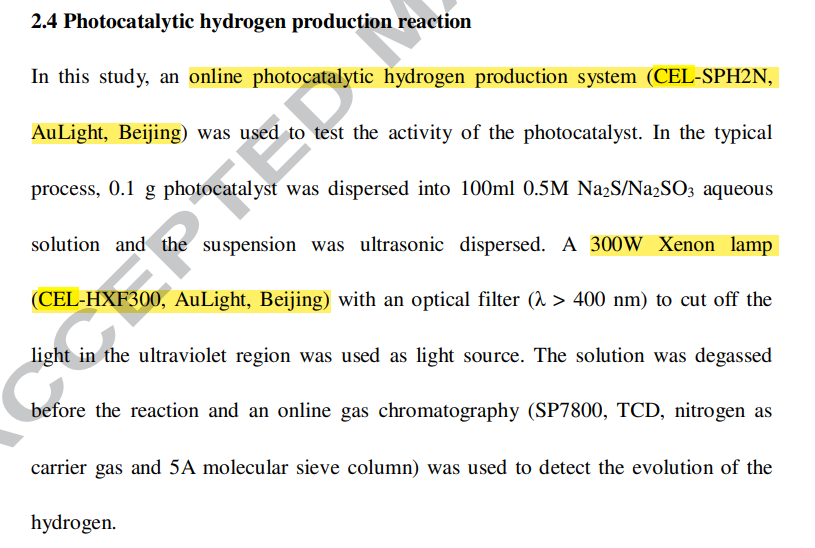
The solution was degassed before the reaction and an online gas chromatography (SP7800, TCD, nitrogen as carrier gas and 5A molecular sieve column) was used to detect the evolution of the hydrogen.
2.5 Characterization Atomic force microscopy (AFM) was performed on a Veeco/DI atomic force microscope instrument. The morphologies were investigated by scanning electron microscopy (SEM, Hitachi, S-3400N) and transmission electron microscopy (TEM, JEOL, JEM-1400). Powder X-ray diffraction (XRD, RIGAKU, D/max2550) was used to characterize the crystalline phase of products. Cu Kα radiation (λ = 0.15405 nm, 40 kV, 100 mA) was used and 2θ scanning range was 10-80°. UV-vis diffuse reflectance spectra (DRS) were obtained with a Scan UV-vis spectrophotometer (Varian, Cary 500) within a range 200-800 nm. Raman spectra measurements were recorded with an inVia Reflex Raman spectrometer with 524.5 nm laser excitation from 100 to 3000 cm -1. The X-ray photoelectron spectra (XPS, Thermo Scientific, ESCALAB 250Xi) were carried out using Al Kα radiation. All binding energies were referred to the C 1s peak of 284.8 eV. Elemental analysis was conducted using Agilent 725ES inductively coupled plasma atomic emission spectrometry (ICP-AES). Photoluminescence spectra were recorded using Fluorolog-3-P (Jobin Yvon).
3. Results and discussion
3.1 Catalyst structure The aqueous dispersion of graphene oxide was prepared through oxidation of graphite flake using KMnO4/H2SO4/H3PO4 according to the literature procedure. Fig. 1 showed the atomic force microscopy (AFM) images of the graphene oxide sheets. The thickness of the graphene sheets was measured as about 1.1 nm, which is in agreement with the literature [24], indicating the formation of the single layered graphene oxide.
The morphology of the organic polymer microspheres, the PSGM/GO and PSGM/rGO/CdS was investigated using SEM, TEM and HRTEM instruments. Fig. 2a shows the SEM image of the original organic polymer microspheres. The synthesized microspheres were micron-sized and its diameter was about 2.5 µm. When the aqueous dispersion of graphene oxide sheets was mixed with the suspension of the amine-functionalized polymer microspheres, the single layered graphene oxide sheets with epoxy groups reacted and formed C-N bond with the amine group on the surface of the polymer microspheres. The graphene oxide sheets were firmly coated on the surface of the microspheres (Fig. 2b) and separated from each other, resulting in less opportunity for agglomeration.
The PSGM/rGO/CdS catalyst was prepared through a solvothermal method. DMSO was used as solvent, sulfur source and reductant of graphene oxide. The temperature of the reaction was set as 453 K and the reaction time was changed from 3 h to 12 h after the reaction temperature increased to 453 K. During the solvothermal process, graphene oxide was reduced and CdS was loaded on the surface of PSGM/rGO microspheres (Fig. 2c). As shown in Fig. 2(e-f), the surface of PSGM/rGO/CdS catalyst was covered with CdS. When the reaction time was set as 3 h, the surface of the microspheres was covered with a thin layer of CdS. It was seen from Fig. 2g that the diameter of CdS particles loaded on the surface of the PSGM/rGO microspheres was about 6-15 nm. When looking into the high resolution HRTEM image of PSGM/rGO/CdS-3 (Fig. 2g), the lattice spacing of 0.336 nm corresponding to (111) facet of cubic phase CdS was observed on the surface of the microsphere, indicating the formation of heterojunction between CdS and rGO. In addition, TEM-EDX measurement was implemented to investigate the chemical compositions at the interface (Fig. 2h). Signals of C, S and Cd elements were observed from the spectra, which further confirmed the formation of heterojunction. As the reaction time increased, the surface of the microspheres covered with more CdS and CdS gradually aggregated to larger particles. It was worth noting that the size of the CdS particles grown on the surface of the microspheres increased with the reaction time and the amount of CdS covered on the surface changed.
To determine the amount of CdS loaded on the surface of the microspheres, ICP-AES measurement was carried out. The percentage of CdS catalyst for the samples synthesized with different reaction time (3 h, 6 h and 12 h) were 15.46 wt%, 35.02 wt% and 39.41 wt%, indicating different loading amount at different reaction time. It was observed that the weight percent of CdS increased with the extension of reaction time. It was deduced that in the initial stage of the synthesis reaction, CdS was produced. As the reaction to proceeded, more CdS was synthesized and loaded on the surface of the PSGM/rGO microspheres. As a result, the weight percent of CdS increased.
Beijing China Education Au-light Co., Ltd.
Room 401, Fengtai Science and technology innovation center, No. 7 Kexing Road, Fengtai District, Beijing
+86 18518316054
eric@aulight.com;info@aulight.com

Scan and pay attention to us